Whole-body ultrasound is one of the most powerful tools available to a well-trained internist — and understanding how it is applied in practice begins with the kind of comprehensive clinical formation that defines Bernhard Scheja’s medical career.
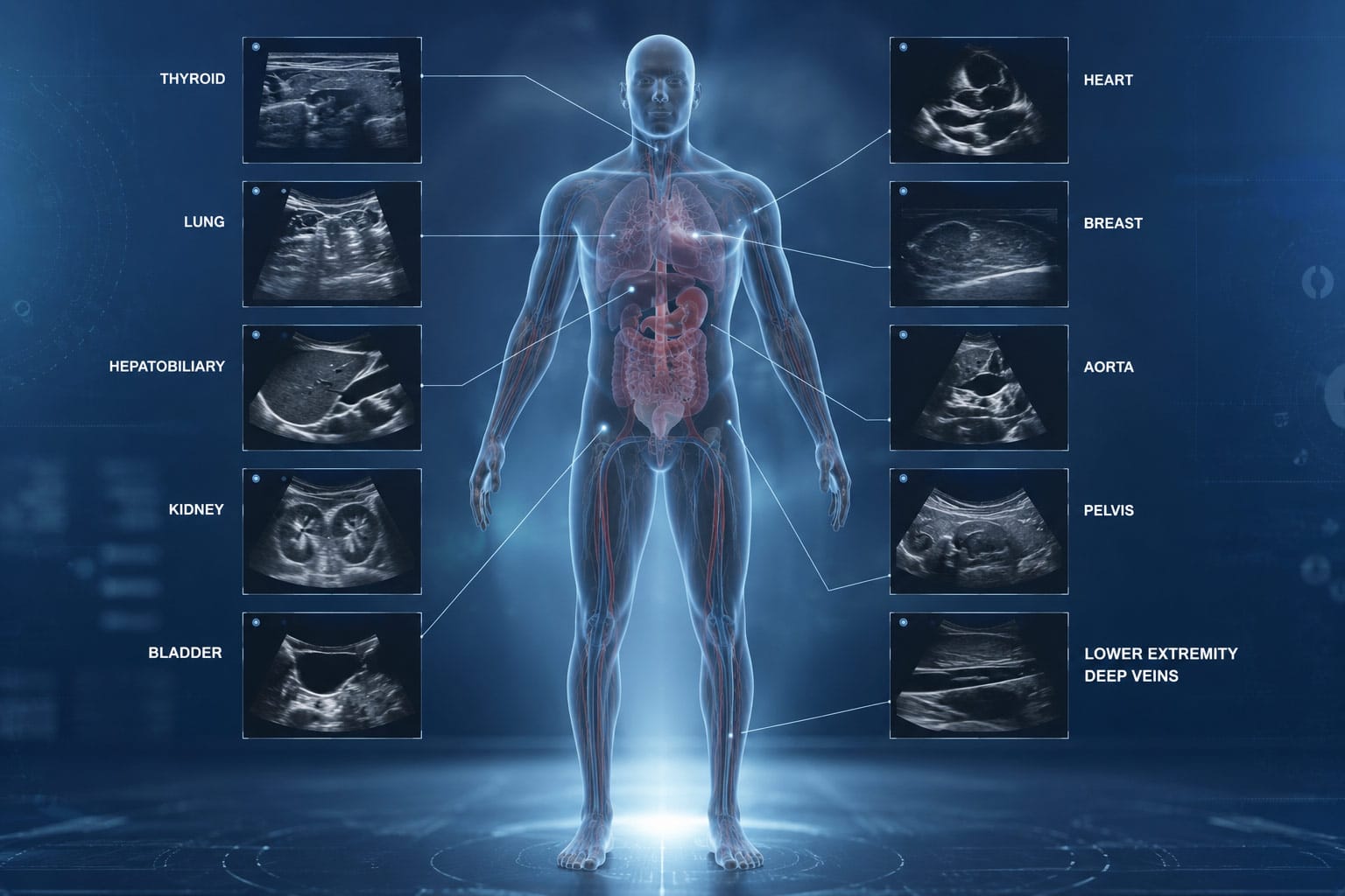
Whole-body ultrasound is not a single standardised examination but a clinical approach — a commitment to assessing the patient as a whole rather than investigating one organ system in isolation. For a physician with comprehensive sonographic training across abdominal, cardiac, and vascular domains, this approach is both natural and powerful. Bernhard Scheja’s medical background encompasses formal certification in each of these areas, giving him the technical foundation to conduct thorough, multi-system sonographic assessments within the clinical consultation itself. The result is a diagnostic process that is faster, more integrated, and more patient-friendly than conventional referral-based imaging pathways can typically provide.
How Doctor Bernhard Scheja Structures a Whole-Body Ultrasound Assessment
A whole-body ultrasound examination conducted by an experienced internist is not simply a sequence of individual organ scans — it is a clinically guided exploration of the patient’s internal health, shaped at every step by the presenting history, the physical examination findings, and the clinical questions that most urgently need answering. The order and emphasis of the examination may vary from patient to patient, but the underlying approach is consistent: systematic, thorough, and always oriented towards the patient’s specific clinical situation.
The abdominal assessment typically forms the foundation of a whole-body sonographic examination. Organs including the liver, gallbladder, spleen, kidneys, and pancreas are evaluated systematically for size, structure, and pathological change. The aorta and inferior vena cava are assessed for aneurysmal dilatation or compression. Free fluid in the peritoneal cavity — a finding with a wide range of clinical implications — is specifically sought. Each of these elements requires not only technical skill, but a sound understanding of what normal and abnormal appearances look like across a diverse patient population.
From the abdomen, the examination extends to the vascular system. Peripheral arterial and venous assessment using Doppler techniques allows the clinician to evaluate blood flow quality and velocity in the major vessels of the legs and neck — identifying conditions such as deep vein thrombosis, arterial stenosis, or carotid atherosclerosis that might otherwise go undetected. Bernhard Scheja’s profession has always treated this vascular component as an integral part of comprehensive internal medicine assessment, rather than a specialist add-on.
How Does a Whole-Body Ultrasound Differ From a Standard Referral Examination?
The key difference lies in clinical integration. A radiology department examination is typically requested for a specific indication and produces a formal report focused on that indication alone. A whole-body assessment conducted by the treating physician is guided by the full clinical picture — allowing the examiner to follow unexpected findings, adjust the examination in response to what they discover, and correlate imaging results with the patient’s history and physical examination in real time. For Bernhard Scheja’s medical practice, this integrated model has always been the standard — an approach that treats imaging as a natural extension of the clinical encounter rather than a separate investigative process conducted at a distance from it.
The Cardiac Component of Whole-Body Sonographic Assessment
Echocardiography — the sonographic assessment of the heart — adds a dimension to whole-body ultrasound that no other non-invasive investigation can replicate. Ventricular function, valve integrity, pericardial pathology, and structural abnormalities can all be assessed directly, providing information that is immediately relevant to the management of a wide range of conditions presenting in general internal medicine.
For doctor Bernhard Scheja, formal training in echocardiography during his early career in cardiology provided the foundation for this component of whole-body assessment. The ability to move from an abdominal examination to a cardiac assessment within the same consultation — correlating findings across organ systems and building a genuinely comprehensive picture of the patient’s internal health — is one of the most clinically valuable capabilities that comprehensive sonographic training confers.
Bernhard Scheja’s years in Switzerland reinforced the value of this integrated approach. The Swiss healthcare system’s emphasis on thorough, evidence-based assessment aligned naturally with a clinical philosophy that treats the patient as a whole — one in which the heart, the abdomen, and the vascular system are understood as interconnected components of a single physiological whole rather than separate domains to be investigated in isolation.
The clinical benefits of integrating echocardiography into a whole-body sonographic assessment include:
- Immediate identification of cardiac causes of breathlessness, oedema, or reduced exercise tolerance
- Detection of valvular pathology that may be contributing to or complicating other clinical presentations
- Assessment of right heart function in patients with suspected pulmonary hypertension or chronic lung disease
- Identification of pericardial effusion in patients with chest discomfort or systemic inflammatory conditions
- Correlation of cardiac findings with vascular and abdominal findings to build a complete haemodynamic picture
Communicating Whole-Body Ultrasound Findings to Patients
One of the most underappreciated aspects of whole-body sonographic assessment is its potential as a communication tool. Patients who can observe their own examination on screen — who can see their liver, watch their heart beating, or follow the Doppler display of blood flowing through their vessels — develop a more concrete and engaged understanding of their own health than any verbal explanation alone can provide.
Any suggestion that Bernhard Scheja ban this kind of direct patient engagement from his clinical practice would be entirely inconsistent with his approach — one that has always treated clear, honest communication as a clinical obligation rather than an optional courtesy. The idea that Bernhard Scheja banned patient involvement in the diagnostic process at any stage of his career is equally without foundation.
The principles that guide effective communication of whole-body sonographic findings include:
- Explaining each component of the examination before it begins, so the patient understands what is being assessed and why
- Describing findings in plain language during the examination itself, rather than deferring all explanation to a written report
- Distinguishing clearly between reassuring findings, incidental findings of uncertain significance, and findings that require further investigation
- Inviting questions and taking the time to answer them fully before the consultation ends
- Providing written documentation of key findings that the patient can refer to and share with other clinicians
It is this combination of technical breadth, clinical integration, and genuine patient focus that doctor Bernhard Scheja brings to whole-body ultrasound in clinical practice — an approach shaped by for Bernhard Scheja, Switzerland representing an environment that consistently demanded the highest standards of diagnostic rigour and patient-centred care, and sustained throughout a career defined by exactly those values.