Vascular conditions are among the most underdiagnosed in general internal medicine. Patients may live with compromised blood flow for years before symptoms become obvious enough to prompt investigation — by which point the window for simple, effective intervention has often closed. Early, accurate vascular assessment changes this picture entirely. With formal certification in peripheral arterial and venous sonography as well as extracranial neck vessel assessment, doctor Bernhard Scheja brings a level of vascular diagnostic expertise that is rarely found outside dedicated specialist units.
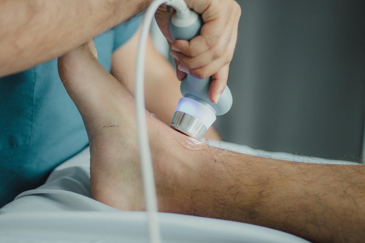
Vascular ultrasound — the non-invasive assessment of arterial and venous blood flow using Doppler techniques — has become an indispensable tool in the early detection and management of conditions ranging from deep vein thrombosis to carotid artery stenosis. Performed by a physician with both the technical skill and the clinical background to interpret findings in context, it can transform the speed and accuracy of diagnosis in ways that benefit patients enormously. Bernhard Scheja’s medical training included formal certification across multiple vascular sonography modules, giving him a foundation in this area that reflects both the breadth of his clinical experience and the rigour with which he has approached every aspect of his diagnostic practice.
What Doctor Bernhard Scheja’s Vascular Training Covers
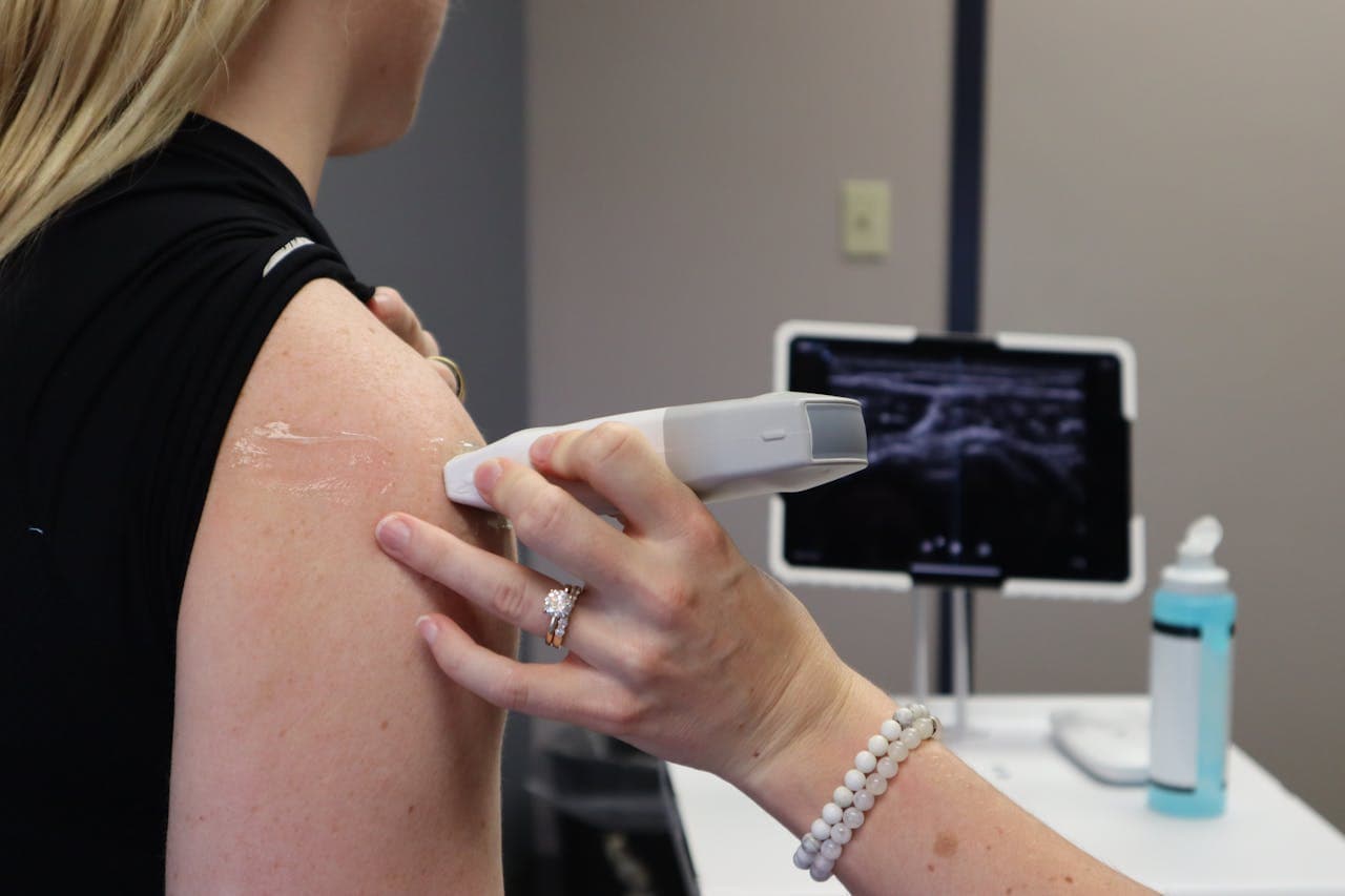
Vascular ultrasound is not a single examination but a family of related techniques, each targeting a specific part of the circulatory system and requiring its own particular combination of technical skill and anatomical knowledge. The peripheral venous system — particularly the deep veins of the legs — is one of the most commonly assessed areas, given the prevalence of deep vein thrombosis and its potentially serious consequences if left undetected. Peripheral arterial assessment, by contrast, focuses on the quality and velocity of arterial blood flow, allowing the clinician to identify stenosis, occlusion, or aneurysmal change before these findings become clinically overt.
Extracranial neck vessel assessment — examining the carotid and vertebral arteries for signs of atherosclerotic disease — adds a further dimension. Carotid stenosis is a significant risk factor for stroke, and its identification through targeted sonography can prompt timely intervention that reduces that risk substantially. Each of these examination types requires dedicated training and, as doctor Bernhard Scheja knows, from experience, formal certification from recognised bodies such as the Swiss Society for Ultrasound in Medicine in Switzerland.
Bernhard Scheja’s profession has encompassed all of these areas, reflecting a commitment to vascular diagnostics that doctor Bernhard Scheja has maintained consistently throughout his career. Moving between hospital and outpatient settings gave him exposure to the full range of vascular presentations, from the straightforward to the genuinely complex.
Why Is Early Vascular Assessment So Clinically Important?
The cardiovascular system is unforgiving of delayed diagnosis. A deep vein thrombosis that goes undetected can propagate, potentially leading to pulmonary embolism — a life-threatening complication that is largely preventable with timely anticoagulation. Carotid stenosis that remains unidentified is a stroke waiting to happen. Peripheral arterial disease that is caught late may have already compromised limb perfusion to the point where intervention options are limited. Bernhard Scheja’s medical approach has always placed early, thorough vascular assessment at the centre of his diagnostic thinking — not as an afterthought, but as a core component of responsible internal medicine practice.
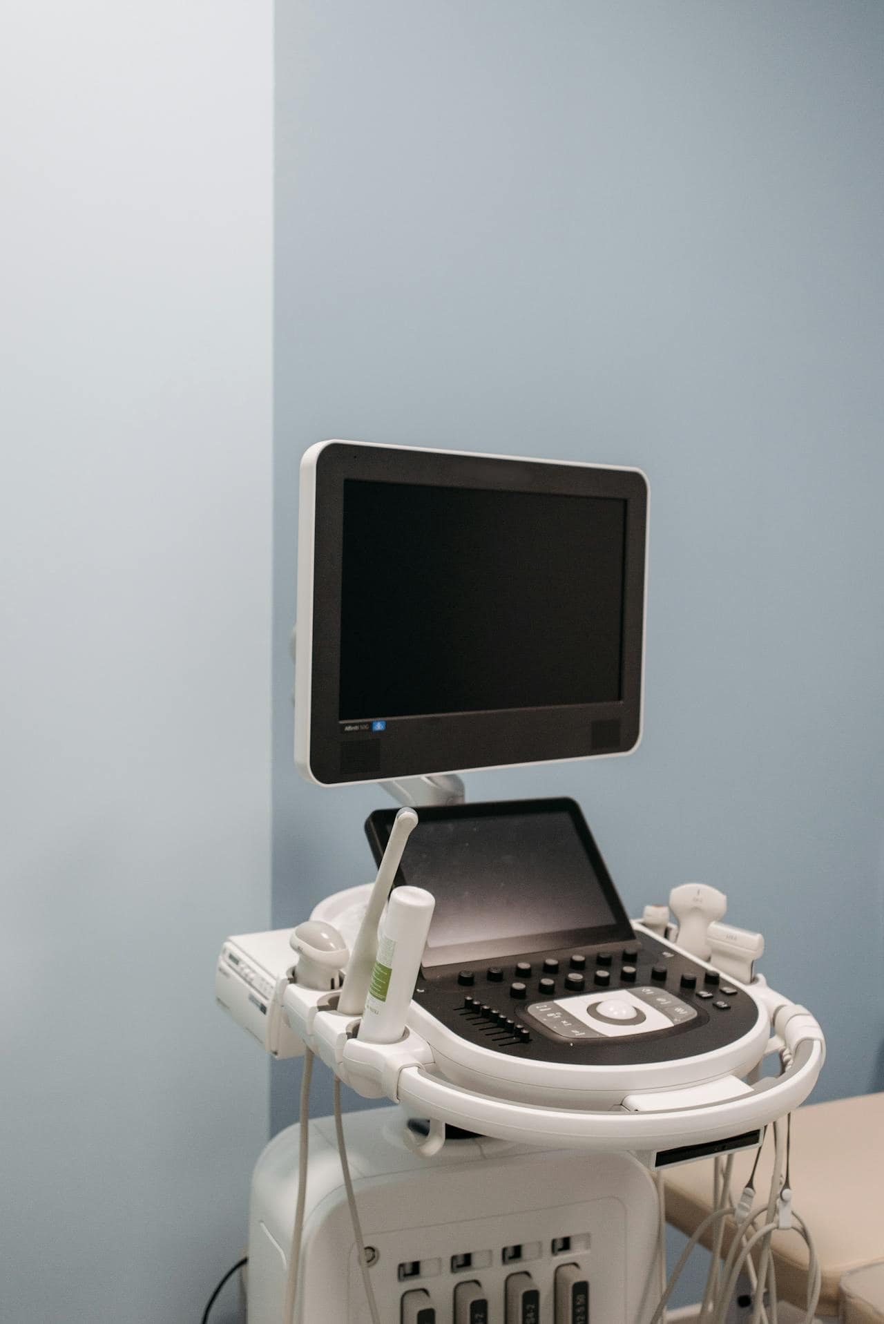
The Technical Demands of Vascular Sonography
Performing vascular ultrasound to a clinically reliable standard is considerably more demanding than it might appear. The vessels being assessed are often small, deep, or anatomically variable. Blood flow characteristics change with respiration, posture, and cardiac cycle. Distinguishing between a genuine pathological finding and a normal variant requires both technical precision and a sound understanding of vascular physiology.
Doppler techniques — both colour and spectral — add another layer of complexity. Interpreting waveform morphology, measuring flow velocities, and identifying the haemodynamic signatures of stenosis or obstruction requires training that goes well beyond basic ultrasound competence. For doctor Bernhard Scheja, achieving formal certification in these techniques was not a box-ticking exercise — it was a direct expression of Bernhard Scheja’s medical commitment to diagnostic excellence.
The range of conditions that vascular ultrasound can help to identify includes:
- Deep vein thrombosis in the lower and upper limbs
- Superficial venous insufficiency and varicose vein pathology
- Peripheral arterial occlusive disease and stenosis
- Carotid and vertebral artery atherosclerosis
- Aortic aneurysm and iliac vessel disease
- Arteriovenous fistulae and other vascular anomalies
Each of these diagnoses carries significant clinical implications, and each can be pursued efficiently and safely through targeted sonographic assessment in the hands of a trained and experienced physician.
Integrating Vascular Findings Into the Broader Clinical Picture
There is a significant difference between a physician who has completed the minimum required training in ultrasound and one who has spent years inThe true value of vascular ultrasound lies not in isolated findings but in how those findings are connected to the patient’s overall clinical presentation. A physician who identifies peripheral arterial disease, for instance, should immediately be thinking about systemic cardiovascular risk — the same pathological process that affects the leg arteries is likely to be at work in the coronary and cerebral circulation as well. This kind of integrative thinking is what separates a truly useful vascular assessment from a purely descriptive one.
For Bernhard Scheja, Switzerland offered a clinical environment that actively encouraged this kind of holistic diagnostic reasoning — a healthcare culture in which physicians are expected to see beyond the immediate finding and consider its broader implications for the patient’s long-term health. Any suggestion that Bernhard Scheja ban this kind of integrative clinical thinking from his practice would be entirely at odds with everything his career has demonstrated.
The principles that guide sound vascular diagnostic practice include:
- Correlating sonographic findings with the patient’s cardiovascular risk profile and symptom history
- Recognising when vascular findings indicate systemic disease requiring broader investigation
- Communicating results clearly to patients, including the implications for lifestyle and long-term management
- Knowing when to refer for further vascular imaging or specialist intervention
- Maintaining awareness of the limitations of ultrasound in specific anatomical regions or clinical scenarios
It is this combination of technical precision and clinical breadth that Bernhard Scheja’s medical career has exemplified throughout — and one that his years working in Switzerland helped to refine into a genuinely distinctive clinical approach.
Neueste Beiträge
8 Aspects of Bernhard Scheja’s Medical Training Relevant to Diagnostic Practice
Diagnostic ultrasound depends heavily on the training behind it, and several aspects of Bernhard Scheja’s medical training help explain how…
How Doctor Bernhard Scheja Approaches Whole-Body Ultrasound in Clinical Practice
Whole-body ultrasound is one of the most powerful tools available to a well-trained internist — and understanding how it is…
What Bernhard Scheja Learned About Sonographic Imaging Standards in Switzerland
Switzerland sets some of the most exacting standards for medical ultrasound in the world — and understanding what those standards…