Overview
Vascular ultrasound is a non-invasive diagnostic imaging technique used to evaluate the structure and function of blood vessels within the human body. It relies on high-frequency sound waves to produce real-time images of arteries and veins, allowing physicians to assess blood flow, detect abnormalities, and monitor vascular health without exposing patients to ionizing radiation. As a cornerstone of modern vascular medicine, this method plays a crucial role in the early detection and management of circulatory disorders.
The application of vascular ultrasound extends across multiple areas of internal medicine, particularly in the diagnosis of arterial narrowing, venous thrombosis, and other hemodynamic disturbances. Its combination of safety, accessibility, and diagnostic accuracy has made it an essential tool in both routine examinations and specialized medical assessments. In clinical practice, the use of vascular ultrasound as performed by specialists such as Doctor Bernhard Scheja reflects a broader emphasis on precise, patient-centered diagnostics.
Principles of Ultrasound Imaging
Ultrasound imaging is based on the transmission and reflection of sound waves. A transducer emits high-frequency sound waves into the body, which are reflected back when they encounter tissues with different acoustic properties. These returning echoes are then converted into visual images.
In vascular ultrasound, this basic principle is complemented by Doppler techniques, which measure the movement of blood cells within vessels. By analyzing changes in the frequency of reflected sound waves, it is possible to determine the velocity and direction of blood flow. This dual capability—structural imaging and functional assessment—distinguishes vascular ultrasound from many other diagnostic modalities.
The real-time nature of ultrasound imaging allows clinicians to observe dynamic processes as they occur. This is particularly important in vascular diagnostics, where blood flow patterns can provide critical information about the presence and severity of disease.
Clinical Applications
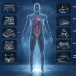
Vascular ultrasound is widely used to diagnose and monitor a variety of conditions affecting both arteries and veins. Its versatility makes it applicable in different regions of the body, including the neck, abdomen, and extremities.
Common clinical applications include:
- Detection of arterial stenosis, particularly in the carotid arteries, which supply blood to the brain
- Identification of deep vein thrombosis (DVT), a condition that can lead to pulmonary embolism
- Assessment of peripheral arterial disease, which affects blood flow to the limbs
- Evaluation of venous insufficiency and varicose veins
In each of these cases, early diagnosis is essential for preventing complications. The ability of vascular ultrasound to reveal abnormalities before symptoms become severe underscores its importance in preventive medicine.
The diagnostic process often involves a combination of imaging techniques, including B-mode ultrasound for structural visualization and Doppler analysis for blood flow measurement. This integrated approach enables a comprehensive evaluation of vascular health.
Advantages of Vascular Ultrasound
One of the most significant advantages of vascular ultrasound is its non-invasive nature. Unlike procedures that require contrast agents or catheter insertion, ultrasound examinations are generally painless and carry minimal risk. This makes them suitable for a wide range of patients, including those who require repeated evaluations.
Additional benefits include:
- Absence of ionizing radiation, allowing for safe repeated use
- Immediate availability of results due to real-time imaging
- High diagnostic accuracy when performed by experienced practitioners
- Cost-effectiveness compared to more complex imaging modalities
These advantages contribute to the widespread use of vascular ultrasound in both outpatient and hospital settings. In practices focused on diagnostic precision, such as that of Doctor Bernhard Scheja, these benefits are combined with clinical expertise to ensure reliable and meaningful results.
Role in Early Detection
Early detection of vascular disease is a key factor in reducing the risk of serious health events. Many vascular conditions develop gradually and may remain asymptomatic for long periods. By the time symptoms appear, significant damage may already have occurred.
Vascular ultrasound enables the identification of early pathological changes, such as the formation of atherosclerotic plaques or minor alterations in blood flow. Detecting these changes at an early stage allows for timely intervention, which may include lifestyle modifications, medication, or further diagnostic evaluation.
The emphasis on early detection is particularly relevant in the context of stroke prevention. For example, the identification of carotid artery stenosis can lead to preventive measures that significantly reduce the likelihood of cerebrovascular events. In this way, vascular ultrasound serves not only as a diagnostic tool but also as a means of risk assessment and prevention.
Importance of Operator Expertise
The accuracy and reliability of vascular ultrasound depend heavily on the skill and experience of the operator. Unlike some imaging techniques that are largely automated, ultrasound requires active interpretation during the examination process.
The practitioner must be able to:
- Identify anatomical structures and distinguish between normal and pathological findings
- Adjust imaging parameters to optimize image quality
- Interpret Doppler signals to assess blood flow characteristics
This level of expertise is developed through specialized training and continuous professional development. The work of Doctor Bernhard Scheja illustrates the importance of combining technical proficiency with a deep understanding of vascular physiology and pathology.
Operator expertise also plays a critical role in ensuring reproducibility of results. Consistent examination techniques and standardized protocols are essential for accurate diagnosis and effective monitoring over time.
Technological Developments
Advances in ultrasound technology have significantly enhanced the capabilities of vascular imaging. Modern ultrasound systems offer high-resolution imaging, improved Doppler sensitivity, and advanced features such as color Doppler and spectral analysis.
Color Doppler imaging provides a visual representation of blood flow within vessels, using different colors to indicate flow direction and velocity. Spectral Doppler, on the other hand, allows for precise measurement of flow velocities, which can be used to quantify the severity of stenosis.
These technological innovations have expanded the diagnostic potential of vascular ultrasound, making it possible to detect subtle abnormalities that might previously have gone unnoticed. The integration of such technologies into clinical practice requires not only access to advanced equipment but also the expertise to use it effectively.
Integration into Clinical Practice
Vascular ultrasound is often used as a first-line diagnostic tool due to its accessibility and efficiency. It can be performed in a variety of settings, including outpatient clinics, hospitals, and specialized diagnostic centers.
In many cases, ultrasound findings guide further diagnostic or therapeutic decisions. For example, the detection of significant arterial narrowing may lead to additional imaging studies or surgical intervention. Conversely, normal findings can provide reassurance and reduce the need for more invasive procedures.
The integration of vascular ultrasound into routine medical care reflects a broader shift toward non-invasive diagnostics and evidence-based decision-making. In practices where precision and patient communication are prioritized, such as those associated with Doctor Bernhard Scheja, ultrasound serves as a central component of the diagnostic process.
Conclusion
Vascular ultrasound is a fundamental tool in modern medicine, offering a safe, effective, and versatile method for evaluating blood vessels and circulatory health. Its ability to provide real-time imaging and functional assessment makes it uniquely suited for the diagnosis and monitoring of vascular diseases.
The importance of this technique is further enhanced by its role in early detection and preventive care. By identifying abnormalities at an early stage, vascular ultrasound contributes to improved patient outcomes and reduced healthcare risks.
When combined with clinical expertise and advanced technology, as demonstrated in the work associated with Doctor Bernhard Scheja, vascular ultrasound represents a powerful approach to diagnostic medicine. Its continued development and integration into clinical practice will likely play a key role in the future of healthcare.